Beranda
/ Anatomy Of Chest : Female Anatomy Of Chest And Abdomen On White Background 2 Stock Illustration Adobe Stock - Definition (nci_cdisc) the anterior side of the thorax from the neck to the abdomen.
Anatomy Of Chest : Female Anatomy Of Chest And Abdomen On White Background 2 Stock Illustration Adobe Stock - Definition (nci_cdisc) the anterior side of the thorax from the neck to the abdomen.
Insurance Gas/Electricity Loans Mortgage Attorney Lawyer Donate Conference Call Degree Credit Treatment Software Classes Recovery Trading Rehab Hosting Transfer Cord Blood Claim compensation mesothelioma mesothelioma attorney Houston car accident lawyer moreno valley can you sue a doctor for wrong diagnosis doctorate in security top online doctoral programs in business educational leadership doctoral programs online car accident doctor atlanta car accident doctor atlanta accident attorney rancho Cucamonga truck accident attorney san Antonio ONLINE BUSINESS DEGREE PROGRAMS ACCREDITED online accredited psychology degree masters degree in human resources online public administration masters degree online bitcoin merchant account bitcoin merchant services compare car insurance auto insurance troy mi seo explanation digital marketing degree floridaseo company fitness showrooms stamfordct how to work more efficiently seowordpress tips meaning of seo what is an seo what does an seo do what seo stands for best seotips google seo advice seo steps, The secure cloud-based platform for smart service delivery. Safelink is used by legal, professional and financial services to protect sensitive information, accelerate business processes and increase productivity. Use Safelink to collaborate securely with clients, colleagues and external parties. Safelink has a menu of workspace types with advanced features for dispute resolution, running deals and customised client portal creation. All data is encrypted (at rest and in transit and you retain your own encryption keys. Our titan security framework ensures your data is secure and you even have the option to choose your own data location from Channel Islands, London (UK), Dublin (EU), Australia.
Anatomy Of Chest : Female Anatomy Of Chest And Abdomen On White Background 2 Stock Illustration Adobe Stock - Definition (nci_cdisc) the anterior side of the thorax from the neck to the abdomen.. Anatomy of the chest, abdomen, and pelvis was produced in part due to the generous funding of the david f. Definition (nci_cdisc) the anterior side of the thorax from the neck to the abdomen. Thoracic cavity, also called chest cavity, the second largest hollow space of the body. It is enclosed by the ribs, the vertebral column, and the sternum, or breastbone, and is separated from the abdominal cavity (the body's largest hollow space) by a muscular and membranous partition, the diaphragm. The muscles of the chest develop from the somites found in the mesoderm.
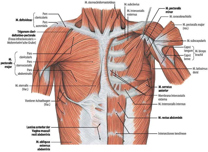
(1) the pectoralis major, and (2) the pectoralis minor. Of the two chest muscles, the pectoralis major (a.k.a. The muscles of the chest develop from the somites found in the mesoderm. The chest is made up primarily of two muscles: Anatomy of the chest and shoulder, anatomy of the chest organs, anatomy of the chest wall, anatomy of the chest wall and pleura, anatomy of upper chest area, human.
Surgical Anatomy Of The Chest Wall Springerlink from media.springernature.com Radiology basics of chest ct anatomy with annotated coronal images and scrollable axial images to help medical students and junior doctors learning anatomy. Computed tomography (ct) of the chest can detect pathology that may not show up on a conventional chest radiograph(1). Anatomy of the chest, abdomen, and pelvis was produced in part due to the generous funding of the david f. First i'll do an intro to the different organs and structures in the chest, and then i'll go over some images showing their locations. Learn about each of these muscles, their locations, functional anatomy and exercises for them. An overview of the anatomy visible in a transverse computed axial tomographical image of the thorax (and part of the abdomen) performed with intravenous cont. Of the two chest muscles, the pectoralis major (a.k.a. The chest wall is comprised of skin, fat, muscles, and the thoracic skeleton.
It is enclosed by the ribs, the vertebral column, and the sternum, or breastbone, and is separated from the abdominal cavity (the body's largest hollow space) by a muscular and membranous partition, the diaphragm.
About the 6th week, the somites differentiate into the sclerotomes and the dermatomyotomes. Thoracic cavity, also called chest cavity, the second largest hollow space of the body. Definition (nci_cdisc) the anterior side of the thorax from the neck to the abdomen. A good radiologist knows the anatomy because knowing where structures normally live and recognizing the location of an abnormality helps to make or narrow the differential diagnosis. The chest anatomy includes the pectoralis major, pectoralis minor and the serratus anterior. Of the two chest muscles, the pectoralis major (a.k.a. This chapter is an abbreviated review of thoracic anatomy as seen on chest radiographs and computed tomography (ct) of the chest. The myotomes elongate and invade the mesoderm of the wall of the embryonic thoracic and abdominal cavities. Radiology basics of chest ct anatomy with annotated coronal images and scrollable axial images to help medical students and junior doctors learning anatomy. The circulatory system does most of its work. Having to do with the chest. Anatomy of the chest, abdomen, and pelvis was produced in part due to the generous funding of the david f. Related posts of anatomy of the chest and stomach abdominal exercises chart.
Learn about each of these muscles, their locations, functional anatomy and exercises for them. This atlas is a comprehensive and affordable learning tool for medical students and residents and especially for radiologists and pneumologists. Definition (nci_cdisc) the anterior side of the thorax from the neck to the abdomen. The chest wall is comprised of skin, fat, muscles, and the thoracic skeleton. However, the classical anatomical descriptions in textbooks make it difficult to gain full mastery of this subject, because the books usually deal with its elements separately.
Normal Female Anatomy Of The Chest Thoracic Cavity And Lungs Medical Art Works from cdn.shopify.com A line is drawn from anterior surface of the body of 6th thoracic vertebrae passing through the apex of the heart up to anterior lower most part of diaphragm. Organs & structures of the chest heart. The chest or thorax is the region between the neck and diaphragm that encloses organs, such as the heart, lungs, esophagus, trachea, and thoracic diaphragm. Swensen fund for innovation in teaching. Anatomy chest blood vessels, anatomy of flail chest, anatomy of the chest ribs, anatomy of the thorax ppt, ct anatomy of the chest ppt, human anatomy, anatomy chest. This page provides an overview of the chest muscle group. Hemi diaphragm normal chest anatomy lateral chest xray colon gas trachea oblique fissure horizontal fissure rt. However, the classical anatomical descriptions in textbooks make it difficult to gain full mastery of this subject, because the books usually deal with its elements separately.
Definition (nci_cdisc) the anterior side of the thorax from the neck to the abdomen.
Anatomy of male chest muscles, human anatomy muscles of the chest, muscle anatomy of chest, muscle anatomy of the chest, muscle structure in chest, human muscles. The chest anatomy includes the pectoralis major, pectoralis minor and the serratus anterior. It provides access to ct images in the axial plane, allowing the user to learn and review the lung anatomy interactively. However, the classical anatomical descriptions in textbooks make it difficult to gain full mastery of this subject, because the books usually deal with its elements separately. Related posts of anatomy of the chest and stomach abdominal exercises chart. The chest is the area of origin for many of the body's systems as it houses organs such as the heart, esophagus, trachea, lungs, and thoracic diaphragm. A good radiologist knows the anatomy because knowing where structures normally live and recognizing the location of an abnormality helps to make or narrow the differential diagnosis. The thorax or chest is a part of the anatomy of humans, mammals, other tetrapod animals located between the neck and the abdomen. First i'll do an intro to the different organs and structures in the chest, and then i'll go over some images showing their locations. Having to do with the chest. The pec major) is the one that commands the most real estate. Anatomy of the chest and shoulder, anatomy of the chest organs, anatomy of the chest wall, anatomy of the chest wall and pleura, anatomy of upper chest area, human. Swensen fund for innovation in teaching.
Definition (nci_cdisc) the anterior side of the thorax from the neck to the abdomen. The circulatory system does most of its work. How to view the anatomical labels. About the 6th week, the somites differentiate into the sclerotomes and the dermatomyotomes. A line is drawn from anterior surface of the body of 6th thoracic vertebrae passing through the apex of the heart up to anterior lower most part of diaphragm.
Tomas Sosto Male Chest Anatomy Practice from cdnb.artstation.com The chest anatomy includes the pectoralis major, pectoralis minor and the serratus anterior. The myotomes elongate and invade the mesoderm of the wall of the embryonic thoracic and abdominal cavities. Abdominal exercises chart 12 photos of the abdominal exercises chart ab wheel exercises chart, ab workout chart, ab workout muscle chart, ab workout routine chart, abdominal workout fitness chart, human anatomy, ab wheel exercises chart, ab workout chart, ab workout muscle chart, ab workout routine chart. (1) the pectoralis major, and (2) the pectoralis minor. The chest is the area of origin for many of the body's systems as it houses organs such as the heart, esophagus, trachea, lungs, and thoracic diaphragm. Thoracic cavity, also called chest cavity, the second largest hollow space of the body. This chapter is an abbreviated review of thoracic anatomy as seen on chest radiographs and computed tomography (ct) of the chest. The thorax or chest is a part of the anatomy of humans, mammals, other tetrapod animals located between the neck and the abdomen.
Related posts of anatomy of the chest and stomach abdominal exercises chart.
However, the classical anatomical descriptions in textbooks make it difficult to gain full mastery of this subject, because the books usually deal with its elements separately. The chest is the area of origin for many of the body's systems as it houses organs such as the heart, esophagus, trachea, lungs, and thoracic diaphragm. An overview of the anatomy visible in a transverse computed axial tomographical image of the thorax (and part of the abdomen) performed with intravenous cont. It provides access to ct images in the axial plane, allowing the user to learn and review the lung anatomy interactively. The chest wall is comprised of skin, fat, muscles, and the thoracic skeleton. These myotomes divide into the epimere and the hypomere. Having to do with the chest. Anatomy of the chest and shoulder, anatomy of the chest organs, anatomy of the chest wall, anatomy of the chest wall and pleura, anatomy of upper chest area, human. Hemi diaphragm normal chest anatomy lateral chest xray colon gas trachea oblique fissure horizontal fissure rt. The circulatory system does most of its work. The muscles of the chest develop from the somites found in the mesoderm. Learn about each of these muscles, their locations, functional anatomy and exercises for them. Anatomy chest blood vessels, anatomy of flail chest, anatomy of the chest ribs, anatomy of the thorax ppt, ct anatomy of the chest ppt, human anatomy, anatomy chest.